There are certain tests and procedures which are considered the "standard of care" and should be performed on every patient at every eye examination. Other tests and procedures, which aren't necessarily classified as "standard of care" should be performed during a complete or "comprehensive" examination in order to ensure that nothing has been missed and there are no conditions or diseases present which have been overlooked. All of these tests and more are part of a "Comprehensive Eye Exam" when you have your eyes examined at the Primary Eye Care Center of Ahwatukee.
There are also numerous other tests and procedures which, depending on your family medical history, your own personal medical history, or your results during standard testing, may be recommended by our doctor. These additional procedures are called "ancillary" tests and are often covered under your medical insurance as opposed to your vision insurance which only covers the standard testing of a comprehensive examination.
Below, we will describe all of the tests which should be performed as part of a thorough and comprehensive eye examination:
Standard Tests in our Eye Examinations:
CASE HISTORY: Any thorough medical examination begins with a complete case history. We review all the relevant information concerning your general health, family history and your past and present medical history. We also review all medications you may be taking including over-the-counter medications which can often affect the eyes. We carefully review any allergies, past optical history, and visual requirements for your occupation as well as any hobbies. Any current visual problems or ocular symptoms are investigated and we check all of the prescriptions in your current eyewear.
COLOR TESTING: Color testing is performed at each examination not only to determine whether a patient is "color blind" or deficient. Reduction in color perception can be an important finding in many eye and neurological diseases including multiple sclerosis. So we test each patient for color perception when performing their annual examination.
BLOOD PRESSURE & PULSE RATE: Hypertension (High Blood Pressure) and heart disease are two of the most common causes of death in the United States. Inside the eye, especially on the retina, there are many blood vessels which supply oxygen and nutrients through the blood stream which are essential to maintaining good vision. Any disruption in the vascular system can lead to loss of vision and even blindness. So we screen patients for hypertension and heart disease and our doctor will check each patient's retinal vasculature for signs of vascular disease at every examination.
STEREOPSIS: A good indication of whether your eyes are working together properly, muscle control, depth perception, and whether you have proper binocular vision can be assertained by steropsis testing. We test and measure the steropsis or "binocular disparity" of each patient during your comprehensive examination.
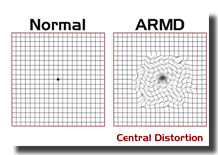
MACULAR FUNCTION: One of the most common causes of vision loss today is "macular degeneration". The macula is the center of your retina and is responsible for achieving good vision. If it degenerates, you lose central vision and cannot read, drive, see television or a computer screen or another person's face. Today, we have treatments for the so called "wet" form of macular degeneration which are very effective and can save a patient's precious central vision if caught early. So during a comprehensive examination, we measure macular function and macular health using several tests.
TONOMETRY: Glaucoma is the second leading cause of new blindness in the United States. In glaucoma patients, the pressure inside the eye becomes too elevated which damages the optic nerve and causes blindness. It is a "silent disease", meaning you will not notice any symptoms until you are about 6 months from being blind. It can be treated with medication to save your vision, but only if caught early. Once your vision has been lost it cannot be restored. We measure the pressure inside your eye using a procedure called "Tonometry". At our office, we have several different types of tonometers to measure the pressure inside your eye. We utilize a screening tonometer also known as an "Air-Puff" tonometer and also use "Applanation Tonometers" which measure the pressure directly without a puff of air. Tonometry is just one of the many tests we perform to detect and treat glaucoma during a comprehensive examination.
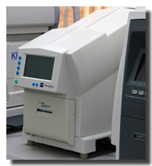
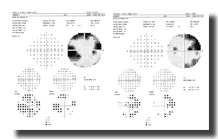
VISUAL FIELDS: Another test which looks for signs of glaucoma is visual field testing which measures your peripheral vision. Visual field mapping can not only detect early stages of glaucoma, but can also detect many neurological conditions as well. Brain tumors, strokes, and multiple sclerosis are a few of the conditions we have uncovered through visual field testing. Often the patient may be asymptomatic and unaware that they have a serious medical problem. By screening for visual field defects during a comprehensive vision examination, we can detect these types of neurological diseases early enough to start treatment. When a screening visual field indicates abnormalities our doctor may order a more thorough visual field called a "Threshold Visual Field" which completely maps out your peripheral visual field thresholds and compares them to a normal database for your age.
BINOCULAR VISION: We test your eye coordination and muscle control to make sure your eyes work together properly. This includes an analysis of the muscles used for movement, focusing, and depth perception. We make sure your eyes are aiming straight at their target and can track objects and scan across a page of words properly. If binocular vision problems are found vision therapy can be recommended or, in some cases, prism or other corrective aids can be prescribed in your glasses as a remedy.
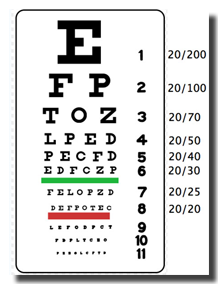
VISUAL ACUITIES: This is a standardized, quantitative measurement of the spatial resolution or "sharpness" of your vision. It is most commonly measured using a "Snellen" eye chart. Several different methods are used to record a patient's visual acuity. In most cases we record the acuity as a fraction such as 20/20 or 20/50 vision. This refers to being able to see a certain size letter at a certain distance. For example, if you can see 20/40 vision this means you are able to see a "40" sized letter at 20 feet distance. This 40 sized letter will be exactly twice the size of a "20" sized letter. So at 20/40 vision you either need to be half the distance or have an object twice the size in order to see what a person with 20/20 can see. 20/20 does not mean perfect vision, it is just a standard we measure your vision against.
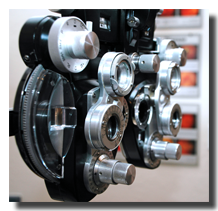
REFRACTION: This is the testing the doctor performs to determine whether you need to wear corrective lenses to improve your vision. Precise measurements are made using several different methods to help the doctor prescribe eyeglasses or contact lenses to give you the best visual acuity possible. We utilize both objective and subjective testing during a refraction. Objective tests may include "Retinoscopy" where the doctor shines a light off the back of your eye (your retina) and neutralizes the reflection using various lenses. This allows the doctor to calculate your prescription without asking you any questions. We also use an "AutoRefractor" which is an automatic computerized version of Retinoscopy. Subjective testing is when the doctor asks you which lens looks better: "number one, or number two?" After taking into consideration all of these tests, the doctor will write a final prescription for your eyeglasses or contact lenses as needed to correct your vision.
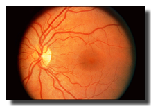
RETINAL EVALUATION: A "Fundus" or Retinal Evaluation is a fundamental part of a complete eye examination. Regularly done as a dilated examination, evaluating the entire retina allows the doctor to check for peripheral retinal problems such as retinal tears, holes, detachments, and various vascular or systemic diseases. The optic nerve is viewed in magnified stereoptic quality or "3D". The macula or center of your retina is evaluated for any signs of degeneration or diseases which may need to be treated. Many retinal problems and diseases can be successfully treated when caught early with either medications or surgery. However, these diseases often have no noticable symptoms until it is too late to restore your vision. So careful examinations on a annual basis are critical to ensuring you maintain your precious vision throughout your lifetime.
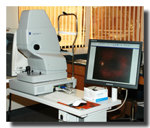
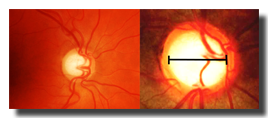
RETINAL PHOTOGRAPHY: In addition to viewing the entire retina, macula, and optic nerve in detail, it is important to document exactly the size and shape of the optic nerve and any abnormalities on the retina. Only by watching for changes can many diseases, such as glaucoma, be diagnosed. We utilize a "Non-Mydriatic" Retinal Camera which allows us to take high definition magnified photographs of the optic nerve, macula, and posterior pole of the retina during every comprehensive eye examination. Even during years when we are not dilating the eye, we can record the appearance of your optic nerve and look for changes from previous exams. In this way, we have the ability to detect diseases such as glaucoma as early as possible so our doctor can begin treatment before vision has been lost.
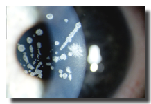
BIOMICROSCOPY: Biomicroscopy, which is also called a "Slit Lamp Examination", is the microscopic examination of living tissues in the eye. Using a microscope called a slit lamp, the doctor can magnify and create a cross section to evaluate individual layers of the tissues in your eye. The cornea can be viewed from front to back. Any opacities or abnormalities can easily be seen in the anterior chamber or in the lens. Cataracts can be evaluated and measured. By using special magnifying lenses in conjunction with the slit lamp, the doctor can also see the posterior chamber and evaluate the vitreous humor as well as the retina and optic nerve in microscopic and three dimensional detail. Contact lenses can be seen and their fit can be evaluated. Any signs of hypoxia or lack of oxygen transmission through the contact lens can be found before vision loss or damage to the cornea occurs. Also, using special stains, the doctor can look for signs of irritation, infections, allergies, and toxins that are affecting your eyes. Biomicroscopy is an important component of a comprehensive eye examination at the Primary Eye Care Center of Ahwatukee.
CORNEAL TOPOGRAPHY: Corneal Topography is a non-invasive medical imaging procedure which maps out the topography of your cornea in 3D. This allows the doctor to view the entire shape of the front surface of your eye and detect any abnormalities or imperfections which may be present. This technique is critical when fitting a contact lens onto the eye to ensure that the lens fits properly and that a contact lens is not causing any damage to the cornea. Topography is also used when evaluating a patient for refractive surgeries such as LASIK or for post-operative examination of surgical procedures. It is also invaluable when working with patients who suffer from corneal diseases such as Keratoconus and patients who have had corneal grafts or transplants. We routinely perform topography as part of a comprehensive eye examination and perform a more extensive version on all patients who are either contact lens wearers or interested in being fit with contact lenses.


Ishihara Color Plates

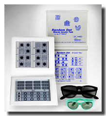
Random Dot Stereopsis

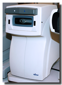

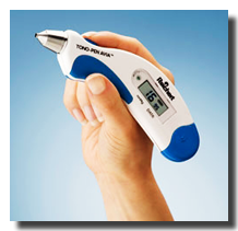
The TonoPen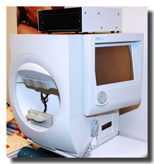




RetinalCross Section


For regularly scheduled eye exams, expect to talk about any changes in your medical history since the last time you saw your eye doctor.
